Global Histology and Cytology Market - Key Trends and Drivers Summarized
Is Histology and Cytology the Foundation of Modern Diagnostic Medicine?
Histology and cytology are integral to the foundation of diagnostic medicine, but why are these fields so crucial for understanding disease at the cellular level? Histology involves the study of tissues under a microscope, allowing pathologists to examine the structure and function of cells within tissues. Cytology, on the other hand, focuses on individual cells or small clusters of cells to detect abnormalities. Together, these disciplines are used to diagnose a wide range of diseases, including cancer, infections, and inflammatory conditions, by revealing microscopic changes in cell and tissue architecture that cannot be seen with the naked eye.The appeal of histology and cytology lies in their ability to provide definitive diagnoses for a variety of conditions, guiding treatment decisions and improving patient outcomes. In oncology, for instance, both disciplines are critical in diagnosing cancers, determining tumor grades, and identifying the spread of malignancies. Additionally, histology and cytology are used in routine screenings, such as Pap smears for cervical cancer and biopsies for breast cancer, playing a pivotal role in early detection and prevention. As personalized medicine and precision healthcare advance, the demand for more accurate, detailed, and rapid diagnostic techniques continues to grow, making histology and cytology even more essential in modern medicine.
How Has Technology Advanced Histology and Cytology?
Technological advancements have significantly transformed histology and cytology, enhancing the speed, accuracy, and scope of diagnostic capabilities. One of the most notable developments has been the introduction of digital pathology and whole-slide imaging. Digital pathology allows entire slides to be scanned at high resolution, enabling pathologists to review tissue samples on computer screens rather than through traditional microscopes. This has revolutionized workflow efficiency, enabling rapid collaboration among pathologists globally and facilitating second opinions or specialized reviews in complex cases. Digital imaging also allows for advanced image analysis using artificial intelligence (AI), which can automatically detect abnormalities, measure tissue components, and highlight areas of interest, improving diagnostic accuracy and consistency.Another significant advancement is the automation of histology and cytology processes. Automated tissue processors, slide staining robots, and cytology specimen preparation machines have streamlined laboratory workflows, reducing human error and improving reproducibility. Automated systems can now handle high volumes of samples while maintaining consistency in staining and preparation, allowing pathologists to focus more on interpretation and diagnosis rather than manual tasks. This has also reduced turnaround times, improving patient care by delivering faster diagnoses.
The development of immunohistochemistry (IHC) and molecular pathology techniques has expanded the diagnostic power of histology. IHC uses antibodies to detect specific proteins within tissue sections, allowing pathologists to identify cancer subtypes, assess hormone receptor status in breast cancer, or detect infectious agents. This molecular-level insight is crucial for personalized medicine, where specific biomarkers guide targeted therapies. Advances in fluorescent in situ hybridization (FISH) and next-generation sequencing (NGS) have also brought molecular diagnostics into the realm of histology and cytology. These techniques allow for the detection of genetic mutations, chromosomal abnormalities, and gene amplifications, providing critical information for the diagnosis and treatment of cancer, genetic disorders, and infectious diseases.
In cytology, advancements in liquid-based cytology (LBC) have greatly improved the quality of cell samples used for analysis. In traditional cytology, such as the Pap smear, cells are manually spread onto slides, which can lead to uneven distribution and air-drying artifacts. LBC, on the other hand, suspends cells in a liquid medium, allowing for a more uniform and representative sample to be prepared. This has led to improved accuracy in detecting precancerous and cancerous changes, especially in cervical cancer screening. LBC has also expanded the range of cytological testing to include tests for human papillomavirus (HPV), which is a significant advancement in cervical cancer prevention.
Another major technological leap is the integration of AI and machine learning in cytology and histology. AI algorithms are being trained to assist in screening and diagnosis by analyzing cell morphology and tissue architecture more rapidly and consistently than human counterparts. In cytology, AI-powered systems are being developed to automatically screen for abnormal cells in samples, such as in Pap smears or fine-needle aspiration biopsies, identifying potential malignancies with high accuracy. In histology, AI can be used to assist in the grading of tumors, detection of metastasis, or identification of rare diseases, significantly reducing the burden on pathologists and improving diagnostic consistency.
Cryosectioning and advanced tissue staining techniques have also enhanced the capabilities of histology. Cryosectioning allows for the rapid freezing of tissues, enabling the preparation of thin tissue sections for immediate examination. This is particularly useful in intraoperative consultations, where surgeons rely on histological results to make real-time decisions during surgery, such as confirming whether all cancerous tissue has been removed. Advanced staining techniques, such as multiplex immunofluorescence, enable the simultaneous visualization of multiple biomarkers in a single tissue section, providing deeper insights into cellular interactions and the tumor microenvironment.
Why Are Histology and Cytology Critical for Modern Medical Diagnostics?
Histology and cytology are critical for modern medical diagnostics because they provide the foundational tools for detecting, diagnosing, and monitoring a wide range of diseases at the cellular and tissue level. In cancer diagnosis, histological examination is the gold standard for confirming malignancy and determining tumor type, grade, and stage. Accurate classification of tumors is essential for guiding treatment decisions, whether it involves surgery, chemotherapy, radiotherapy, or targeted therapies. By analyzing the architecture of tissues and identifying key cellular changes, histology allows for precise characterization of cancerous growths, including whether they are invasive, metastatic, or benign.In cytology, the ability to evaluate individual cells or small clusters of cells provides valuable insights for early detection and diagnosis of diseases. The Pap smear, a cytological test, is one of the most successful screening tools in medical history, drastically reducing the incidence and mortality of cervical cancer by detecting precancerous cells before they develop into invasive cancer. Cytology is also used in fine-needle aspiration (FNA) biopsies, a minimally invasive technique for sampling suspicious lumps or masses in organs such as the thyroid, breast, or lymph nodes. FNA cytology provides rapid, reliable diagnoses for conditions ranging from cancer to infections and autoimmune diseases, reducing the need for more invasive surgical biopsies.
Histology and cytology are indispensable in diagnosing infectious diseases by identifying pathogens directly in tissue samples or cytological preparations. Histopathology allows for the detection of viral, bacterial, fungal, or parasitic infections, often providing more definitive diagnoses than serological or culture-based methods. In conditions such as tuberculosis, histology can reveal granulomatous inflammation, while special stains may be used to visualize acid-fast bacilli. In viral infections, cytological changes, such as multinucleated giant cells or inclusion bodies, can provide immediate clues to the causative virus. This ability to diagnose infections at a cellular level is particularly important in cases where rapid, targeted treatment is essential, such as in immunocompromised patients or in the management of outbreaks.
In addition to oncology and infectious disease diagnosis, histology and cytology are vital for understanding autoimmune and inflammatory conditions. For instance, in gastrointestinal diseases like Crohn's disease or ulcerative colitis, histological analysis of biopsy samples is used to confirm the diagnosis and assess the severity of inflammation, helping guide treatment plans. Similarly, in conditions such as rheumatoid arthritis, histopathological examination of synovial tissue can reveal characteristic inflammatory patterns, allowing for earlier diagnosis and intervention. By providing detailed information about tissue and cellular changes, histology and cytology allow clinicians to better understand the underlying pathology of diseases, improving diagnosis, prognosis, and treatment decisions.
The role of histology in surgical pathology is also critical during surgeries, particularly in oncology. Surgeons often rely on histological assessments during procedures to ensure that all cancerous tissue has been removed or to determine whether a mass is malignant. This intraoperative consultation, often called a “frozen section,” allows surgeons to make real-time decisions that directly impact the success of the surgery and the patient's prognosis.
Histology and cytology also play a pivotal role in precision medicine. In recent years, the integration of molecular diagnostics with traditional histopathology has enabled a more detailed understanding of diseases at the genetic and molecular levels. By combining molecular data with histological findings, clinicians can tailor treatments to the individual patient based on the specific genetic mutations or molecular characteristics of their disease. For example, in cancer treatment, molecular markers identified through histology, such as HER2 expression in breast cancer or PD-L1 in lung cancer, are used to determine eligibility for targeted therapies or immunotherapies. This personalized approach to treatment improves outcomes by ensuring that patients receive the most effective therapy based on the unique characteristics of their disease.
What Factors Are Driving the Growth of the Histology and Cytology Market?
The growth of the histology and cytology market is driven by several key factors, including the increasing prevalence of cancer and chronic diseases, advancements in diagnostic technologies, rising demand for early detection, and the growing importance of personalized medicine. One of the primary drivers is the rising global incidence of cancer. As the number of cancer cases continues to climb, particularly in aging populations, the need for accurate, rapid, and cost-effective diagnostic tools has grown. Histology and cytology are essential for diagnosing and classifying cancers, making them critical components of the oncology diagnostics market. The demand for biopsies and cytological evaluations is expected to rise in parallel with the increasing prevalence of cancer, driving the growth of histology and cytology services worldwide.Advancements in diagnostic technology are also propelling the histology and cytology market forward. The introduction of digital pathology, AI, and automated systems has significantly improved the efficiency and accuracy of these disciplines, making them more accessible to a broader range of healthcare facilities. Automated slide staining, digital imaging, and AI-powered diagnostic tools have reduced the burden on pathologists, allowing for faster diagnoses and greater consistency across laboratories. These technologies are particularly beneficial in high-volume settings, such as cancer centers or large hospitals, where the ability to process and analyze a high number of samples quickly is essential for maintaining patient care standards.
The rising demand for early disease detection is another key factor driving market growth. In fields such as cancer screening and infectious disease diagnosis, early detection through cytology and histology can drastically improve patient outcomes by enabling earlier intervention. For example, cytological tests like the Pap smear and liquid-based cytology have revolutionized cervical cancer screening, leading to earlier detection of precancerous lesions and reduced mortality rates. As public health campaigns continue to emphasize the importance of regular cancer screenings and early diagnosis of chronic diseases, the demand for histology and cytology services is expected to increase.
Personalized medicine is also contributing to the growth of the histology and cytology market. As more treatments, particularly in oncology, are tailored to the genetic and molecular characteristics of individual patients, the need for detailed diagnostic information has expanded. Histology and cytology provide the foundation for these personalized treatments by offering insights into tissue structure, cellular changes, and biomarker expression. The integration of molecular diagnostics, such as immunohistochemistry, FISH, and NGS, into routine histology and cytology workflows has further enhanced the value of these disciplines in personalized care, driving demand for advanced diagnostic services.
The growing awareness and acceptance of minimally invasive diagnostic techniques, such as fine-needle aspiration cytology (FNAC), are also boosting the cytology market. FNAC provides a less invasive alternative to surgical biopsies, offering rapid and accurate diagnoses for conditions such as thyroid nodules, breast lumps, and lymphadenopathy. The rising preference for minimally invasive procedures among patients and healthcare providers is expected to increase the demand for cytology services, particularly as the technique continues to demonstrate high accuracy and low complication rates.
The expansion of histology and cytology in emerging markets is another driver of growth. As healthcare systems in developing regions improve and access to diagnostic services expands, the demand for histology and cytology is expected to rise. Government initiatives to improve cancer screening programs and infectious disease diagnosis are driving investment in pathology infrastructure, further supporting market growth. In these regions, the adoption of newer technologies, such as digital pathology and automated slide processing, is expected to enhance the availability and quality of histology and cytology services.
With advancements in diagnostic technologies, the rising incidence of cancer and chronic diseases, and the growing focus on personalized medicine, the histology and cytology market is poised for significant growth. As healthcare systems continue to prioritize early detection and precision diagnostics, histology and cytology will remain at the forefront of modern medicine, providing critical insights into disease mechanisms, guiding treatment decisions, and improving patient outcomes across the globe.
Report Scope
The report analyzes the Histology and Cytology market, presented in terms of market value (USD). The analysis covers the key segments and geographic regions outlined below.- Segments: Type of Examination (Cytology, Histology); Product Type (Reagents & Consumables, Instruments & Analysis Software Systems); Application (Drug Discovery & Designing, Clinical Diagnostics, Research).
- Geographic Regions/Countries: World; United States; Canada; Japan; China; Europe (France; Germany; Italy; United Kingdom; and Rest of Europe); Asia-Pacific; Rest of World.
Key Insights:
- Market Growth: Understand the significant growth trajectory of the Cytology segment, which is expected to reach US$50.6 Billion by 2030 with a CAGR of 15.3%. The Histology segment is also set to grow at 12.3% CAGR over the analysis period.
- Regional Analysis: Gain insights into the U.S. market, valued at $8.6 Billion in 2024, and China, forecasted to grow at an impressive 13.5% CAGR to reach $10.5 Billion by 2030. Discover growth trends in other key regions, including Japan, Canada, Germany, and the Asia-Pacific.
Why You Should Buy This Report:
- Detailed Market Analysis: Access a thorough analysis of the Global Histology and Cytology Market, covering all major geographic regions and market segments.
- Competitive Insights: Get an overview of the competitive landscape, including the market presence of major players across different geographies.
- Future Trends and Drivers: Understand the key trends and drivers shaping the future of the Global Histology and Cytology Market.
- Actionable Insights: Benefit from actionable insights that can help you identify new revenue opportunities and make strategic business decisions.
Key Questions Answered:
- How is the Global Histology and Cytology Market expected to evolve by 2030?
- What are the main drivers and restraints affecting the market?
- Which market segments will grow the most over the forecast period?
- How will market shares for different regions and segments change by 2030?
- Who are the leading players in the market, and what are their prospects?
Report Features:
- Comprehensive Market Data: Independent analysis of annual sales and market forecasts in US$ Million from 2024 to 2030.
- In-Depth Regional Analysis: Detailed insights into key markets, including the U.S., China, Japan, Canada, Europe, Asia-Pacific, Latin America, Middle East, and Africa.
- Company Profiles: Coverage of players such as Abbott Laboratories, Becton, Dickinson and Company, Danaher Corporation, F. Hoffmann-La Roche AG, Hologic, Inc. and more.
- Complimentary Updates: Receive free report updates for one year to keep you informed of the latest market developments.
Some of the 36 companies featured in this Histology and Cytology market report include:
- Abbott Laboratories
- Becton, Dickinson and Company
- Danaher Corporation
- F. Hoffmann-La Roche AG
- Hologic, Inc.
- Merck KgaA
- PerkinElmer, Inc.
- Sysmex Corporation
- Thermo Fisher Scientific, Inc.
- Trivitron Healthcare
This edition integrates the latest global trade and economic shifts into comprehensive market analysis. Key updates include:
- Tariff and Trade Impact: Insights into global tariff negotiations across 180+ countries, with analysis of supply chain turbulence, sourcing disruptions, and geographic realignment. Special focus on 2025 as a pivotal year for trade tensions, including updated perspectives on the Trump-era tariffs.
- Adjusted Forecasts and Analytics: Revised global and regional market forecasts through 2030, incorporating tariff effects, economic uncertainty, and structural changes in globalization. Includes historical analysis from 2015 to 2023.
- Strategic Market Dynamics: Evaluation of revised market prospects, regional outlooks, and key economic indicators such as population and urbanization trends.
- Innovation & Technology Trends: Latest developments in product and process innovation, emerging technologies, and key industry drivers shaping the competitive landscape.
- Competitive Intelligence: Updated global market share estimates for 2025, competitive positioning of major players (Strong/Active/Niche/Trivial), and refined focus on leading global brands and core players.
- Expert Insight & Commentary: Strategic analysis from economists, trade experts, and domain specialists to contextualize market shifts and identify emerging opportunities.
Table of Contents
Companies Mentioned (Partial List)
A selection of companies mentioned in this report includes, but is not limited to:
- Abbott Laboratories
- Becton, Dickinson and Company
- Danaher Corporation
- F. Hoffmann-La Roche AG
- Hologic, Inc.
- Merck KgaA
- PerkinElmer, Inc.
- Sysmex Corporation
- Thermo Fisher Scientific, Inc.
- Trivitron Healthcare
Table Information
| Report Attribute | Details |
|---|---|
| No. of Pages | 191 |
| Published | February 2026 |
| Forecast Period | 2024 - 2030 |
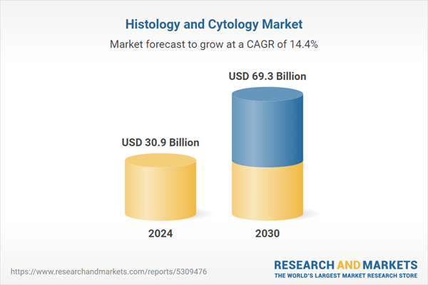
| Estimated Market Value ( USD | $ 30.9 Billion |
| Forecasted Market Value ( USD | $ 69.3 Billion |
| Compound Annual Growth Rate | 14.4% |
| Regions Covered | Global |


