+353-1-416-8900REST OF WORLD
+44-20-3973-8888REST OF WORLD
1-917-300-0470EAST COAST U.S
1-800-526-8630U.S. (TOLL FREE)

Live Cell Imaging Market - Global Industry Size, Share, Trends, Opportunity, & Forecast, 2019-2029F

  • PDF Icon

    Report

  • 185 Pages
  • May 2024
  • Region: Global
  • TechSci Research
  • ID: 5972919
Free Webex Call
10% Free customization
Free Webex Call

Speak directly to the analyst to clarify any post sales queries you may have.

10% Free customization

This report comes with 10% free customization, enabling you to add data that meets your specific business needs.

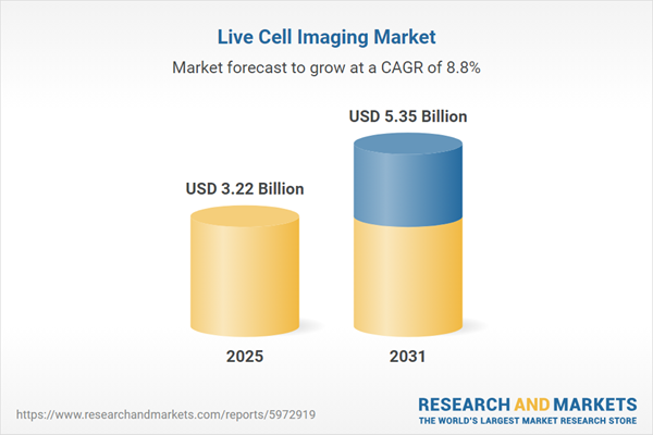
Global Live Cell Imaging Market was valued at USD 2.86 billion in 2023 and will see an robust growth in the forecast period at a CAGR of 11.04% through 2029. Live cell imaging is a scientific technique that allows researchers to observe and analyze living cells in real-time using microscopy and imaging technologies. Unlike traditional fixed-cell imaging, which involves the fixation and staining of cells for static analysis, live cell imaging enables the dynamic visualization of cellular processes, behaviors, and interactions as they occur within living organisms or in vitro cell cultures. Live cell imaging typically involves the use of specialized microscopes equipped with sensitive detectors, high-resolution objectives, and imaging software optimized for capturing dynamic cellular events. Various microscopy techniques, including widefield microscopy, confocal microscopy, multiphoton microscopy, and super-resolution microscopy, can be employed for live cell imaging, each offering unique advantages in terms of spatial resolution, imaging depth, and contrast enhancement. Fluorescent probes, dyes, and genetically encoded markers are commonly used to label specific cellular structures, proteins, organelles, and biomolecules for visualization in live cell imaging experiments. Fluorescent labeling enables researchers to selectively highlight and track cellular components of interest, such as nuclei, cytoskeleton, mitochondria, endoplasmic reticulum, and membrane receptors, facilitating the study of cellular dynamics and functions in real-time. Live cell imaging experiments are performed using specialized cell culture systems and imaging chambers that maintain optimal physiological conditions for cell growth, viability, and function. Temperature-controlled incubators, humidified environments, and CO2-regulated atmospheres ensure stable conditions for cell survival and imaging stability during prolonged experiments.

The growing need for high-resolution imaging techniques to study complex cellular processes and dynamic interactions drives the demand for live cell imaging systems. Researchers and healthcare professionals seek advanced imaging technologies capable of capturing detailed, real-time images of living cells with high spatial and temporal resolution. Live cell imaging plays a crucial role in drug discovery and development by facilitating the real-time monitoring of cellular responses to drug candidates, assessing drug efficacy, and evaluating potential toxicity. The pharmaceutical industry increasingly relies on live cell imaging systems to accelerate the drug development pipeline, reduce costs, and improve the success rate of new therapeutic interventions. Continuous advancements in microscopy techniques, imaging sensors, and analysis software enhance the capabilities and performance of live cell imaging systems. Innovations in microscopy technology enable researchers to capture high-resolution images of living cells with improved sensitivity, speed, and precision, driving market adoption and expansion.

Key Market Drivers

Increasing Demand for High-Resolution Imaging Techniques

High-resolution imaging techniques enable researchers to visualize intricate details of cellular structures and dynamic processes in real-time. Live cell imaging allows for the observation of cellular events such as cell division, migration, signaling, and interaction with high clarity and precision. Live cell imaging provides valuable insights into the functional behavior of cells under various physiological and pathological conditions. High-resolution imaging enables researchers to study subcellular structures, organelle dynamics, and molecular interactions within living cells, contributing to a deeper understanding of cellular function and regulation. High-resolution live cell imaging is instrumental in studying the mechanisms underlying various diseases, including cancer, neurodegenerative disorders, infectious diseases, and metabolic syndromes. Researchers use live cell imaging to investigate disease progression, identify disease biomarkers, and develop targeted therapeutic interventions. High-resolution live cell imaging plays a crucial role in drug discovery and development by facilitating the screening, evaluation, and optimization of potential drug candidates. Researchers use live cell imaging assays to assess drug efficacy, toxicity, and pharmacokinetics in physiologically relevant cellular models, accelerating the drug development pipeline and improving drug safety profiles.

High-resolution live cell imaging allows researchers to monitor cellular responses to external stimuli, environmental cues, and therapeutic interventions in real-time. By visualizing cellular dynamics at high resolution, researchers can track changes in cell morphology, behavior, and function, enabling precise quantification and analysis of cellular responses under dynamic conditions. Continuous advancements in microscopy techniques, imaging sensors, and analysis software enhance the spatial and temporal resolution of live cell imaging systems. High-resolution imaging technologies such as confocal microscopy, multi-photon microscopy, and super-resolution microscopy enable researchers to achieve subcellular-level resolution and capture dynamic cellular events with unprecedented detail and clarity. High-resolution live cell imaging is increasingly being applied in translational research and clinical diagnostics to visualize disease processes, monitor treatment responses, and predict patient outcomes. By integrating high-resolution imaging techniques with clinical workflows, researchers and clinicians can gain insights into disease pathology, identify prognostic markers, and personalize treatment strategies for improved patient care. This factor will help in the development of the Global Live Cell Imaging Market.

Expanding Applications in Drug Discovery and Development

Live cell imaging allows researchers to visualize and monitor cellular responses to drug candidates in real-time. By observing how cells react to various compounds, researchers can assess drug efficacy, toxicity, and mechanisms of action more accurately than with traditional endpoint assays. Live cell imaging systems can be integrated with automated platforms to conduct high-throughput screening of large compound libraries. This enables researchers to rapidly identify lead compounds with desired biological activities and optimize drug candidates for further development. Live cell imaging helps identify novel drug targets by studying cellular processes involved in disease pathology. By observing dynamic changes in cell morphology, proliferation, and function, researchers can pinpoint key signaling pathways and molecular targets for therapeutic intervention. Live cell imaging enables researchers to characterize the mechanisms of action of drugs at the cellular level. By tracking drug interactions with target molecules, observing changes in cellular morphology, and monitoring intracellular signaling pathways, researchers can elucidate how drugs exert their effects and optimize treatment regimens.

Live cell imaging allows for the prediction of drug response based on individual cellular phenotypes and genetic profiles. By analyzing how different cell types or patient-derived cells respond to drug treatments, researchers can tailor therapies to specific patient populations, improving treatment outcomes and minimizing adverse effects. Live cell imaging helps researchers study mechanisms of drug resistance in diseases such as cancer. By observing how cancer cells adapt and develop resistance to chemotherapy or targeted therapies, researchers can identify new strategies to overcome resistance and enhance treatment efficacy. Live cell imaging provides valuable insights into the validity and relevance of preclinical models for drug discovery. By comparing cellular responses observed in live cell imaging assays with clinical outcomes, researchers can validate preclinical models, identify biomarkers of drug response, and improve the translatability of preclinical findings to human disease. This factor will pace up the demand of the Global Live Cell Imaging Market.

Technological Advancements in Microscopy and Imaging Software

Advances in microscopy technology enable the capture of high-resolution images with improved spatial and temporal resolution. Higher resolution allows researchers to visualize cellular structures and dynamic processes at finer detail and track rapid changes in real-time, enhancing the accuracy and reliability of live cell imaging experiments. Super-resolution microscopy techniques, such as structured illumination microscopy (SIM), stimulated emission depletion microscopy (STED), and single-molecule localization microscopy (SMLM), push the limits of optical resolution beyond the diffraction limit. These techniques enable researchers to achieve subcellular-level resolution and visualize molecular structures and interactions with unprecedented clarity and precision. Advanced live cell imaging systems integrate multiple imaging modalities, such as fluorescence, brightfield, phase contrast, and differential interference contrast (DIC) microscopy, to provide complementary information about cellular morphology, dynamics, and function. Multi-modal imaging enhances the versatility and capabilities of live cell imaging platforms, enabling researchers to study a wide range of biological phenomena with greater depth and insight. Continuous improvements in imaging sensors, detectors, and light sources enhance the speed and sensitivity of live cell imaging systems. Faster imaging speeds enable researchers to capture rapid cellular events and dynamic processes with minimal motion blur, while increased sensitivity improves the detection of weak fluorescent signals and reduces phototoxicity and photobleaching effects.

Light sheet microscopy, also known as selective plane illumination microscopy (SPIM), facilitates high-speed, high-resolution imaging of large specimens and three-dimensional (3D) cellular structures. Light sheet microscopy minimizes photodamage to live cells by illuminating only the focal plane of interest, enabling long-term, non-invasive imaging of dynamic biological processes in living organisms and tissue samples. Sophisticated imaging software and analysis tools enable researchers to process, analyze, and visualize large volumes of live cell imaging data with greater efficiency and accuracy. Advanced algorithms for image segmentation, feature extraction, and quantification facilitate automated image analysis, object tracking, and data mining, accelerating the interpretation and extraction of meaningful insights from live cell imaging experiments. Integration of AI and ML algorithms into live cell imaging systems enhances automated image analysis and data interpretation capabilities. AI-based approaches enable the identification of complex cellular phenotypes, classification of cellular events, and prediction of drug responses based on large-scale imaging datasets, empowering researchers to extract valuable biological insights and discover novel patterns and correlations within live cell imaging data. This factor will accelerate the demand of the Global Live Cell Imaging Market

Key Market Challenges

Phototoxicity and Photobleaching

Phototoxicity refers to the harmful effects of light exposure on living cells during imaging experiments. Prolonged or intense illumination can induce cellular stress, DNA damage, and cell death, compromising the integrity of experimental results and affecting the viability and behavior of cells under observation. Phototoxicity can introduce artifacts and distortions in live cell imaging data, leading to inaccurate interpretation and analysis of cellular processes. Photodamaged cells may exhibit abnormal morphology, altered physiological responses, and impaired function, confounding experimental observations and undermining the reliability of experimental outcomes. Phototoxicity can reduce cell viability and compromise the physiological relevance of live cell imaging experiments. Photodamaged cells may undergo apoptosis, necrosis, or senescence, limiting the duration and quality of imaging studies and impeding the study of long-term cellular dynamics and behaviors. Photobleaching refers to the irreversible loss of fluorescence intensity in fluorescently labeled molecules due to repeated light exposure. Photobleaching limits the duration and quality of live cell imaging experiments, as fluorescent signals diminish over time, reducing signal-to-noise ratios and impairing the detection and quantification of cellular structures and processes. Photobleaching reduces the brightness and contrast of fluorescent signals, compromising image quality and resolution in live cell imaging experiments. Diminished fluorescence intensity makes it challenging to distinguish between specific and nonspecific signals, complicating image analysis and interpretation and limiting the sensitivity and accuracy of experimental results. Phototoxicity and photobleaching impose constraints on the duration and frequency of live cell imaging experiments. Researchers must balance the need for high-quality imaging data with the risk of cellular damage and photobleaching effects, optimizing imaging parameters and experimental conditions to minimize adverse effects on cell viability and imaging outcomes.

Standardization and Reproducibility

Live cell imaging experiments are susceptible to variability in experimental conditions, including cell culture protocols, imaging techniques, equipment settings, and environmental factors. Minor variations in these parameters can significantly impact imaging outcomes and experimental results, leading to inconsistencies and difficulties in replicating findings across different research studies. The absence of standardized protocols and guidelines for live cell imaging contributes to variability and inconsistency in experimental procedures and data interpretation. Researchers may use different imaging platforms, software tools, and analytical methods, making it challenging to compare results and reproduce experimental findings across laboratories and research groups. Live cell imaging experiments may be conducted using a wide range of microscopy systems, cameras, objectives, filters, and imaging software, each with its own specifications and performance characteristics. Variations in imaging setups and equipment configurations can introduce biases and errors in imaging data, hindering the reproducibility and reliability of experimental results. Inconsistent sample preparation techniques, cell culture conditions, and handling procedures can affect cell viability, morphology, and behavior during live cell imaging experiments. Variability in sample preparation and handling practices can lead to differences in cellular responses and imaging outcomes, making it challenging to replicate experimental conditions and validate findings across studies. Image analysis in live cell imaging experiments often involves subjective judgments and manual interventions, which can introduce bias and variability in data interpretation. Differences in image processing algorithms, segmentation methods, and quantification criteria can lead to inconsistencies in image analysis and result interpretation, affecting the reproducibility and reliability of experimental findings. Publication bias and selective reporting of positive results in scientific literature can contribute to challenges in reproducibility and data interpretation in live cell imaging studies. Negative or inconclusive findings may be underrepresented or omitted from publications, leading to overestimation of experimental outcomes and limited transparency in reporting experimental methods and results.

Key Market Trends

Emergence of Organ-on-Chip (OOC) and Microfluidics Technologies

Organ-on-Chip (OOC) and Microfluidics platforms allow researchers to replicate complex physiological microenvironments and tissue architectures in vitro. These platforms feature microfluidic channels, chambers, and scaffolds that mimic the spatial organization, mechanical cues, and biochemical gradients present in living tissues, enabling more physiologically relevant cell culture and imaging experiments. Organ-on-Chip (OOC) and Microfluidics technologies are integrated with live cell imaging systems to facilitate real-time visualization and analysis of cellular responses within micro engineered tissue models. Live cell imaging enables researchers to monitor cell behavior, migration, differentiation, and interactions within microfluidic devices, providing insights into dynamic cellular processes and tissue responses under controlled experimental conditions. Organ-on-Chip (OOC) and Microfluidics platforms enable high-throughput screening of drug candidates and therapeutic compounds using live cell imaging assays. By culturing cells in miniature tissue models within microfluidic devices, researchers can screen large libraries of compounds, monitor drug responses, and assess pharmacological effects on cellular physiology and function, accelerating drug discovery and development efforts. Organ-on-Chip (OOC) and Microfluidics technologies support long-term monitoring of cell behavior and responses to dynamic microenvironmental cues. Continuous perfusion of cell culture media, precise control of fluid flow rates, and automated imaging systems allow researchers to sustain cell viability, maintain homeostasis, and monitor cellular dynamics over extended periods, facilitating the study of chronic diseases, tissue regeneration, and developmental processes. Organ-on-Chip (OOC) and Microfluidics platforms integrate multi-modal imaging techniques, such as fluorescence microscopy, confocal microscopy, and live-cell imaging, to capture comprehensive information about cellular morphology, function, and molecular signaling within micro engineered tissues. Multi-modal imaging enables researchers to visualize spatial and temporal changes in cellular behavior, analyze subcellular structures, and investigate molecular interactions in real-time, enhancing the depth and resolution of live cell imaging experiments.

Segmental Insights

Technology Insights

The Time Lapse Microscopy segment is projected to experience rapid growth in the Global Live Cell Imaging Market during the forecast period. Time lapse microscopy allows researchers to capture real-time images of cellular processes, such as cell division, migration, and interactions, over extended periods. This dynamic visualization provides valuable insights into the behavior and dynamics of living cells, enabling the study of biological phenomena with high temporal resolution. Time lapse microscopy plays a crucial role in drug discovery and development by facilitating the real-time monitoring of cellular responses to drug candidates and environmental stimuli. Researchers use time-lapse imaging to assess drug efficacy, toxicity, and pharmacokinetics, accelerating the drug screening process and improving the selection of lead compounds for further development. Time lapse microscopy enables the study of dynamic cellular processes, including cell motility, signaling pathways, and morphological changes, in response to various stimuli and experimental conditions. This longitudinal analysis provides comprehensive insights into the underlying mechanisms governing cellular behavior and function, advancing our understanding of complex biological systems. The integration of three-dimensional (3D) and four-dimensional (4D) imaging techniques into time-lapse microscopy allows researchers to capture spatial and temporal dynamics of cellular structures and interactions in three dimensions. These advanced imaging modalities enhance the spatial resolution and depth perception of time-lapse imaging, enabling more accurate reconstruction and analysis of complex biological processes. Time lapse microscopy is widely adopted across various fields of life sciences research, including cell biology, developmental biology, neuroscience, and cancer research. Its versatility and applicability to a wide range of biological questions make it a valuable tool for studying fundamental biological processes and disease mechanisms.

End-Users Insights

The Pharmaceutical and Biotechnology Companies segment is projected to experience rapid growth in the Global Live Cell Imaging Market during the forecast period. Pharmaceutical and biotechnology companies heavily rely on live cell imaging technologies to accelerate drug discovery and development processes. Live cell imaging enables real-time visualization and analysis of cellular responses to drug candidates, helping researchers identify promising leads, assess drug efficacy, and evaluate potential side effects more efficiently. Live cell imaging systems can be integrated with automated imaging platforms to conduct high-throughput screening of compound libraries for drug discovery purposes. Pharmaceutical and biotechnology companies utilize live cell imaging assays to screen large numbers of compounds, identify hits, and prioritize lead candidates for further optimization and preclinical testing. The shift towards personalized medicine and targeted therapies has increased the demand for live cell imaging technologies in pharmaceutical and biotechnology research. Live cell imaging allows for the characterization of patient-derived cells and tissues, enabling researchers to tailor treatment strategies based on individual genetic profiles and disease phenotypes. Live cell imaging plays a crucial role in stem cell research and regenerative medicine, areas of significant interest for pharmaceutical and biotechnology companies. These companies utilize live cell imaging to study stem cell differentiation, proliferation, and tissue regeneration processes, with the aim of developing novel cell-based therapies for a wide range of diseases and conditions. Continuous advancements in live cell imaging technology, including improvements in microscopy techniques, imaging sensors, and analysis software, have enhanced the capabilities and accessibility of live cell imaging systems for pharmaceutical and biotechnology applications. These technological advancements enable researchers to capture high-resolution, real-time images of cellular processes with greater accuracy and precision.

Regional Insights

North America emerged as the dominant region in the Global Live Cell Imaging Market in 2023. North America boasts a robust infrastructure for biomedical research, including state-of-the-art laboratories, academic institutions, and research centers. The region is home to leading universities, medical schools, and biotechnology companies that drive innovation and technological advancements in live cell imaging. The healthcare and pharmaceutical sectors in North America are highly developed and invest significantly in research and development. The demand for live cell imaging technologies is fueled by the need for advanced tools and techniques to study disease mechanisms, drug interactions, and cellular processes, driving market growth in the region. North American companies and research institutions actively engage in collaborations and partnerships to accelerate product development, innovation, and commercialization of live cell imaging technologies. These collaborations foster knowledge exchange, access to funding, and the development of novel applications, enhancing the competitiveness of the region in the global market. The regulatory environment in North America is conducive to the development and commercialization of life science technologies, including live cell imaging systems. Regulatory agencies such as the FDA (Food and Drug Administration) provide clear guidelines and pathways for product approval, ensuring safety, efficacy, and quality standards are met.

Key Market Players

  • Bio-Rad Laboratories, Inc.
  • Agilent Technologies Inc.
  • Blue-Ray Biotech Corp.
  • CytoSMART Technologies (Axion BioSystems, Inc)
  • Curiosis Inc.
  • Carl Zeiss AG
  • Thermo Fisher Scientific Inc.
  • Perkin Elmer Inc
  • Danaher Corporation
  • Nikon Corporation

Report Scope:

In this report, the Global Live Cell Imaging Market has been segmented into the following categories, in addition to the industry trends which have also been detailed below:

Live Cell Imaging Market, By Product:

  • Instruments
  • Consumables
  • Software
  • Services

Live Cell Imaging Market, By Application:

  • Cell Biology
  • Stem Cells
  • Developmental Biology
  • Drug Discovery

Live Cell Imaging Market, By Technology:

  • Time Lapse Microscopy
  • Fluorescence Recovery After Photo Bleaching
  • High Content Screening
  • Fluorescence Resonance Energy Transfer
  • Others

Live Cell Imaging Market, By End-Users:

  • Pharmaceutical and Biotechnology Companies
  • Academic and Research Institutes
  • Contract Research Organizations

Live Cell Imaging Market, By Region:

  • North America
  • United States
  • Canada
  • Mexico
  • Europe
  • Germany
  • United Kingdom
  • France
  • Italy
  • Spain
  • Asia-Pacific
  • China
  • Japan
  • India
  • Australia
  • South Korea
  • South America
  • Brazil
  • Argentina
  • Colombia
  • Middle East & Africa
  • South Africa
  • Saudi Arabia
  • UAE

Competitive Landscape

Company Profiles: Detailed analysis of the major companies present in the Global Live Cell Imaging Market.

Available Customizations:

Global Live Cell Imaging market report with the given market data, the publisher offers customizations according to a company's specific needs. The following customization options are available for the report:

Company Information

  • Detailed analysis and profiling of additional market players (up to five).

This product will be delivered within 1-3 business days.

Table of Contents

1. Product Overview
1.1. Market Definition
1.2. Scope of the Market
1.2.1. Markets Covered
1.2.2. Years Considered for Study
1.2.3. Key Market Segmentations
2. Research Methodology
2.1. Objective of the Study
2.2. Baseline Methodology
2.3. Key Industry Partners
2.4. Major Association and Secondary Sources
2.5. Forecasting Methodology
2.6. Data Triangulation & Validation
2.7. Assumptions and Limitations
3. Executive Summary
3.1. Overview of the Market
3.2. Overview of Key Market Segmentations
3.3. Overview of Key Market Players
3.4. Overview of Key Regions/Countries
3.5. Overview of Market Drivers, Challenges, Trends
4. Voice of Customer
5. Global Live Cell Imaging Market Outlook
5.1. Market Size & Forecast
5.1.1. By Value
5.2. Market Share & Forecast
5.2.1. By Product (Instruments, Consumables, Software, Services)
5.2.2. By Application (Cell Biology, Stem Cells, Developmental Biology, Drug Discovery)
5.2.3. By Technology (Time Lapse Microscopy, Fluorescence Recovery After Photo Bleaching, High Content Screening, Fluorescence Resonance Energy Transfer, Others)
5.2.4. By End-Users (Pharmaceutical and Biotechnology Companies, Academic and Research Institutes, Contract Research Organizations)
5.2.5. By Region
5.2.6. By Company (2023)
5.3. Market Map
6. North America Live Cell Imaging Market Outlook
6.1. Market Size & Forecast
6.1.1. By Value
6.2. Market Share & Forecast
6.2.1. By Product
6.2.2. By Application
6.2.3. By Technology
6.2.4. By End-Users
6.2.5. By Country
6.3. North America: Country Analysis
6.3.1. United States Live Cell Imaging Market Outlook
6.3.1.1. Market Size & Forecast
6.3.1.1.1. By Value
6.3.1.2. Market Share & Forecast
6.3.1.2.1. By Product
6.3.1.2.2. By Application
6.3.1.2.3. By Technology
6.3.1.2.4. By End-Users
6.3.2. Canada Live Cell Imaging Market Outlook
6.3.2.1. Market Size & Forecast
6.3.2.1.1. By Value
6.3.2.2. Market Share & Forecast
6.3.2.2.1. By Product
6.3.2.2.2. By Application
6.3.2.2.3. By Technology
6.3.2.2.4. By End-Users
6.3.3. Mexico Live Cell Imaging Market Outlook
6.3.3.1. Market Size & Forecast
6.3.3.1.1. By Value
6.3.3.2. Market Share & Forecast
6.3.3.2.1. By Product
6.3.3.2.2. By Application
6.3.3.2.3. By Technology
6.3.3.2.4. By End-Users
7. Europe Live Cell Imaging Market Outlook
7.1. Market Size & Forecast
7.1.1. By Value
7.2. Market Share & Forecast
7.2.1. By Product
7.2.2. By Application
7.2.3. By Technology
7.2.4. By End-Users
7.2.5. By Country
7.3. Europe: Country Analysis
7.3.1. Germany Live Cell Imaging Market Outlook
7.3.1.1. Market Size & Forecast
7.3.1.1.1. By Value
7.3.1.2. Market Share & Forecast
7.3.1.2.1. By Product
7.3.1.2.2. By Application
7.3.1.2.3. By Technology
7.3.1.2.4. By End-Users
7.3.2. United Kingdom Live Cell Imaging Market Outlook
7.3.2.1. Market Size & Forecast
7.3.2.1.1. By Value
7.3.2.2. Market Share & Forecast
7.3.2.2.1. By Product
7.3.2.2.2. By Application
7.3.2.2.3. By Technology
7.3.2.2.4. By End-Users
7.3.3. Italy Live Cell Imaging Market Outlook
7.3.3.1. Market Size & Forecast
7.3.3.1.1. By Value
7.3.3.2. Market Share & Forecast
7.3.3.2.1. By Product
7.3.3.2.2. By Application
7.3.3.2.3. By Technology
7.3.3.2.4. By End-Users
7.3.4. France Live Cell Imaging Market Outlook
7.3.4.1. Market Size & Forecast
7.3.4.1.1. By Value
7.3.4.2. Market Share & Forecast
7.3.4.2.1. By Product
7.3.4.2.2. By Application
7.3.4.2.3. By Technology
7.3.4.2.4. By End-Users
7.3.5. Spain Live Cell Imaging Market Outlook
7.3.5.1. Market Size & Forecast
7.3.5.1.1. By Value
7.3.5.2. Market Share & Forecast
7.3.5.2.1. By Product
7.3.5.2.2. By Application
7.3.5.2.3. By Technology
7.3.5.2.4. By End-Users
8. Asia-Pacific Live Cell Imaging Market Outlook
8.1. Market Size & Forecast
8.1.1. By Value
8.2. Market Share & Forecast
8.2.1. By Product
8.2.2. By Application
8.2.3. By Technology
8.2.4. By End-Users
8.2.5. By Country
8.3. Asia-Pacific: Country Analysis
8.3.1. China Live Cell Imaging Market Outlook
8.3.1.1. Market Size & Forecast
8.3.1.1.1. By Value
8.3.1.2. Market Share & Forecast
8.3.1.2.1. By Product
8.3.1.2.2. By Application
8.3.1.2.3. By Technology
8.3.1.2.4. By End-Users
8.3.2. India Live Cell Imaging Market Outlook
8.3.2.1. Market Size & Forecast
8.3.2.1.1. By Value
8.3.2.2. Market Share & Forecast
8.3.2.2.1. By Product
8.3.2.2.2. By Application
8.3.2.2.3. By Technology
8.3.2.2.4. By End-Users
8.3.3. Japan Live Cell Imaging Market Outlook
8.3.3.1. Market Size & Forecast
8.3.3.1.1. By Value
8.3.3.2. Market Share & Forecast
8.3.3.2.1. By Product
8.3.3.2.2. By Application
8.3.3.2.3. By Technology
8.3.3.2.4. By End-Users
8.3.4. South Korea Live Cell Imaging Market Outlook
8.3.4.1. Market Size & Forecast
8.3.4.1.1. By Value
8.3.4.2. Market Share & Forecast
8.3.4.2.1. By Product
8.3.4.2.2. By Application
8.3.4.2.3. By Technology
8.3.4.2.4. By End-Users
8.3.5. Australia Live Cell Imaging Market Outlook
8.3.5.1. Market Size & Forecast
8.3.5.1.1. By Value
8.3.5.2. Market Share & Forecast
8.3.5.2.1. By Product
8.3.5.2.2. By Application
8.3.5.2.3. By Technology
8.3.5.2.4. By End-Users
9. South America Live Cell Imaging Market Outlook
9.1. Market Size & Forecast
9.1.1. By Value
9.2. Market Share & Forecast
9.2.1. By Product
9.2.2. By Application
9.2.3. By Technology
9.2.4. By End-Users
9.2.5. By Country
9.3. South America: Country Analysis
9.3.1. Brazil Live Cell Imaging Market Outlook
9.3.1.1. Market Size & Forecast
9.3.1.1.1. By Value
9.3.1.2. Market Share & Forecast
9.3.1.2.1. By Product
9.3.1.2.2. By Application
9.3.1.2.3. By Technology
9.3.1.2.4. By End-Users
9.3.2. Argentina Live Cell Imaging Market Outlook
9.3.2.1. Market Size & Forecast
9.3.2.1.1. By Value
9.3.2.2. Market Share & Forecast
9.3.2.2.1. By Product
9.3.2.2.2. By Application
9.3.2.2.3. By Technology
9.3.2.2.4. By End-Users
9.3.3. Colombia Live Cell Imaging Market Outlook
9.3.3.1. Market Size & Forecast
9.3.3.1.1. By Value
9.3.3.2. Market Share & Forecast
9.3.3.2.1. By Product
9.3.3.2.2. By Application
9.3.3.2.3. By Technology
9.3.3.2.4. By End-Users
10. Middle East and Africa Live Cell Imaging Market Outlook
10.1. Market Size & Forecast
10.1.1. By Value
10.2. Market Share & Forecast
10.2.1. By Product
10.2.2. By Application
10.2.3. By Technology
10.2.4. By End-Users
10.2.5. By Country
10.3. MEA: Country Analysis
10.3.1. South Africa Live Cell Imaging Market Outlook
10.3.1.1. Market Size & Forecast
10.3.1.1.1. By Value
10.3.1.2. Market Share & Forecast
10.3.1.2.1. By Product
10.3.1.2.2. By Application
10.3.1.2.3. By Technology
10.3.1.2.4. By End-Users
10.3.2. Saudi Arabia Live Cell Imaging Market Outlook
10.3.2.1. Market Size & Forecast
10.3.2.1.1. By Value
10.3.2.2. Market Share & Forecast
10.3.2.2.1. By Product
10.3.2.2.2. By Application
10.3.2.2.3. By Technology
10.3.2.2.4. By End-Users
10.3.3. UAE Live Cell Imaging Market Outlook
10.3.3.1. Market Size & Forecast
10.3.3.1.1. By Value
10.3.3.2. Market Share & Forecast
10.3.3.2.1. By Product
10.3.3.2.2. By Application
10.3.3.2.3. By Technology
10.3.3.2.4. By End-Users
11. Market Dynamics
11.1. Drivers
11.2. Challenges
12. Market Trends & Developments
12.1. Merger & Acquisition (If Any)
12.2. Product Launches (If Any)
12.3. Recent Developments
13. Porter’s Five Forces Analysis
13.1. Competition in the Industry
13.2. Potential of New Entrants
13.3. Power of Suppliers
13.4. Power of Customers
13.5. Threat of Substitute Product
14. Competitive Landscape
14.1. Bio-Rad Laboratories, Inc.
14.1.1. Business Overview
14.1.2. Company Snapshot
14.1.3. Products & Services
14.1.4. Financials (As Reported)
14.1.5. Recent Developments
14.1.6. Key Personnel Details
14.1.7. SWOT Analysis
14.2. Agilent Technologies Inc.
14.3. Blue-Ray Biotech Corp.
14.4. CytoSMART Technologies (Axion BioSystems, Inc)
14.5. Curiosis Inc.
14.6. Carl Zeiss AG
14.7. Thermo Fisher Scientific Inc.
14.8. Perkin Elmer Inc.
14.9. Danaher Corporation
14.10.Nikon Corporation
15. Strategic Recommendations16. About the Publisher & Disclaimer

Companies Mentioned

  • Bio-Rad Laboratories, Inc.
  • Agilent Technologies Inc.
  • Blue-Ray Biotech Corp.
  • CytoSMART Technologies (Axion BioSystems, Inc)
  • Curiosis Inc.
  • Carl Zeiss AG
  • Thermo Fisher Scientific Inc.
  • Perkin Elmer Inc
  • Danaher Corporation
  • Nikon Corporation

Table Information